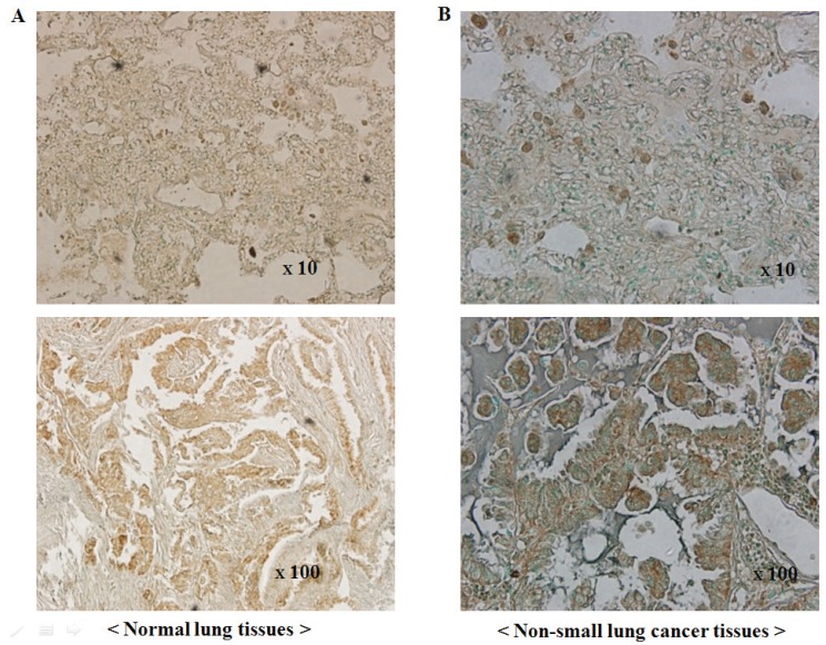
Figure 2.
PLD1 immunohistochemistry in human non-small cell lung cancers and normal lung tissues. (A) Immunoreactivity for PLD1 in normal alveolar lung tissues. The acinar epithelia and intra-alveolar connective tissue exhibit a negative reaction and positive staining, respectively; (B) Immunoreactivity for PLD1 in non-small cell lung cancer tissues (NSCLC). NSCLC tissues exhibit a strong positive reaction (top: ×10, bottom: ×100).

