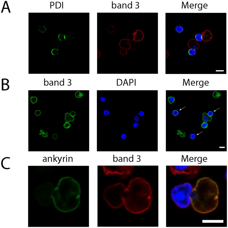
Figure 4. Immunofluorescence microscopy confirms that band 3 and ER are lost during nuclear extrusion.
Human orthochromatic erythroblasts undergoing enucleation after 144 h of differentiation were removed from culture, fixed in 0.5% acrolein and permeabilised using 0.05% Triton X-100. A) Confocal section of enucleating cells labelled with PDI and band 3 antibodies. B) Confocal section showing extruded nuclei (marked with arrows) labelled with band 3 (BRIC170). C) Confocal section of an erythroblast in the late stage of enucleation co-labelled with ankyrin and band 3 antibodies. The non-association of band 3 with ankyrin was observed in every cell identified at the late stage of enucleation (n = 10). Scale bar represents 5 µm.

