
Figure 4. Immune responsive and IMD pathway –controlled splicing factors.
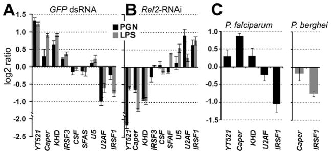
qRT-PCR –based differential transcript abundance analysis of 9 putative splicing factors in the Sua5B cells upon peptidoglycan (PGN) and lipopolysaccharide (LPS) challenge compared to naïve controls (A), or upon Rel2 depletion of PGN and LPS challenged cells compared to GFP dsRNA treated controls (B). (C) qRT-PCR-based differential transcript abundance analysis of 5 or 2 putative splicing factors in P. falciparum infected (left) or P. berghei infected (right) total female mosquitoes compared to naïve controls. Three biological replicates were included and data are represented as mean +/− SEM. See also Table S4.