
Figure 5. Pulsed field gel electrophoresis (PFGE) and Southern blot hybridization results.
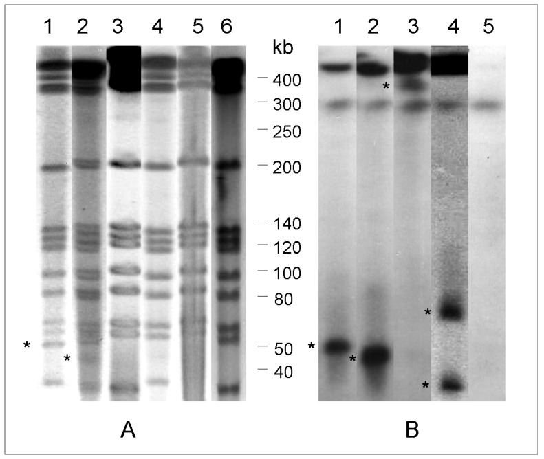
(A) PFGE-DraI and (B) Southern blot hybridization using the trfA-derived probe of selected isolates showing the PFGE patterns of the epidemic strain: 1, the sequenced isolate INCQS 00594, showing the ∼50 bp PFGE-DraI band that hybridized with the trfA derived probe; 2, isolate showing a band with faster migration in PFGE-DraI and Southern blot hybridization; 3, no plasmid band detected in PFGE-DraI and a hybridization band visible near gel origin; 4, two plasmid bands with different migration; 5, no evidence of the presence of pMAB01 either using PFGE-DraI or hybridization; and 6, cured INCQS 00594 colony. The asterisks indicate the plasmid bands.