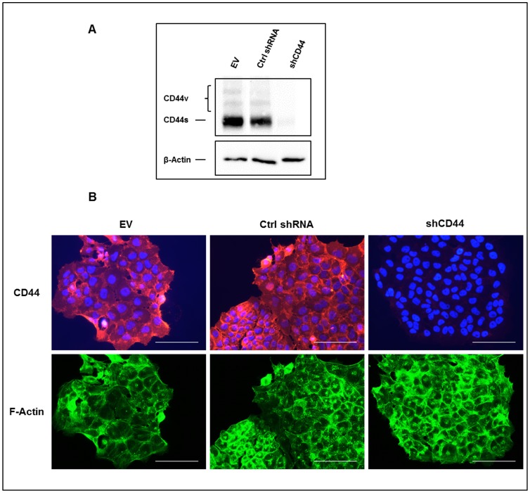
Figure 1. shRNA-mediated downregulation of CD44 expression in 143-B OS cells.
(A) Western blot analysis with the panCD44 Hermes3 antibody of total CD44 gene-derived protein products in extracts of 143-B EV (EV), 143-B Ctrl shRNA (Ctrl shRNA) or 143-B shCD44 (shCD44) cells. β-Actin was used as a loading control. (B) Cell immunostaining of CD44 (red) in saponin permeabilized 143-B EV (EV), 143-B Ctrl shRNA (Ctrl shRNA) or 143-B shCD44 (shCD44) cells. Actin filaments (green) and cell nuclei (blue) were visualized with Alexa Fluor 488-labeled phalloidin and DAPI, respectively. Bars, 100 µm.

