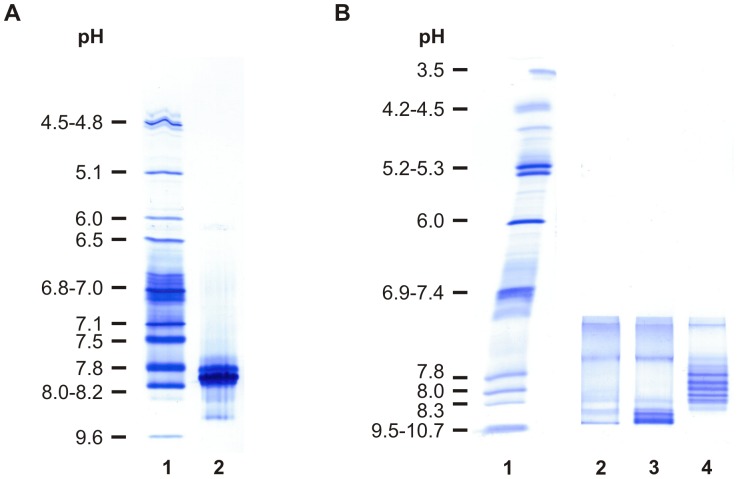
Figure 4. Isoelectric focusing of chCE7agl and chCE7agl-[(DOTA)n-decalysine]2 on an IEF agarose plate pH 3–10.
Samples with a concentration of 5 µg/µL were dialyzed against deionized water before loading and 5 µL of each probe were loaded 4 cm from the cathode. After prefocusing at 1 W constant power for 10 min and focusing for 90 min (20 W power limit; 1000 voltage limit) the gels were fixed in 30% TCA and proteins were stained with Coomassie. A. Lane 1: IEF standard Biorad No 161-0310; lane 2: The parent antibody chCE7agl revealed a pI of 7.8. B. Lane 1: IEF marker Serva No 39212.01. The (DOTA)n-decalysine conjugates showing anodal movement depending on the numbers of DOTA chelats coupled to the decalysine. Lane 2: chCE7agl-[(DOTA)-decalysine]2; lane 3: chCE7agl-[(DOTA)3-decalysine]2; lane 4: chCE7agl-[(DOTA)5-decalysine]2.

