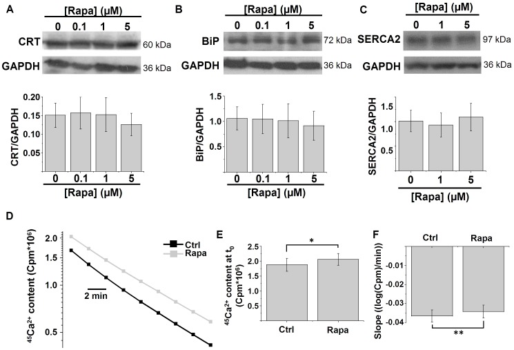
Figure 3. Rapamycin reduces the ER Ca2+-leak rate.
A–B) Western-blot analysis for luminal Ca2+-binding proteins in HeLa cells treated with the indicated concentrations of rapamycin (Rapa) for 5 h: calreticulin (CRT) (A) and BiP/Grp78 (BiP) (B). Upper panels: representative Western blots; lower panels: quantification of the protein/GAPDH ratio (n = 4). C) Western-blot analysis for SERCA2 in HeLa cells treated with the indicated concentrations of rapamycin for 5 h. Upper panels: representative Western blots; lower panel: quantification of the SERCA2/GAPDH ratio (n = 4). D) Representative plot showing the decrease in ER 45Ca2+ content (logarithmic scale) in a Ca2+-free efflux medium without ATP as a function of time in permeabilized HeLa cells pretreated for 5 h with 1 µM rapamycin or with DMSO. The passively bound Ca2+ was determined by loading the cells with 45Ca2+ in the presence of 10 µM of the Ca2+ ionophore A23187 and then subtracted from the stored 45Ca2+. The ER Ca2+-leak rate can be estimated as the rate of decline of the ER 45Ca2+-store content as a function of time. E) Quantification of the mean 45Ca2+-store content at the beginning of the measurement (t0) (n = 5). F) Quantification of the mean slope of the curve in D after transformation to a linear scale, which is a measure of the 45Ca2+-leak rate (n = 5). * p<0.05; ** p<0.01, paired Student's t-test.

