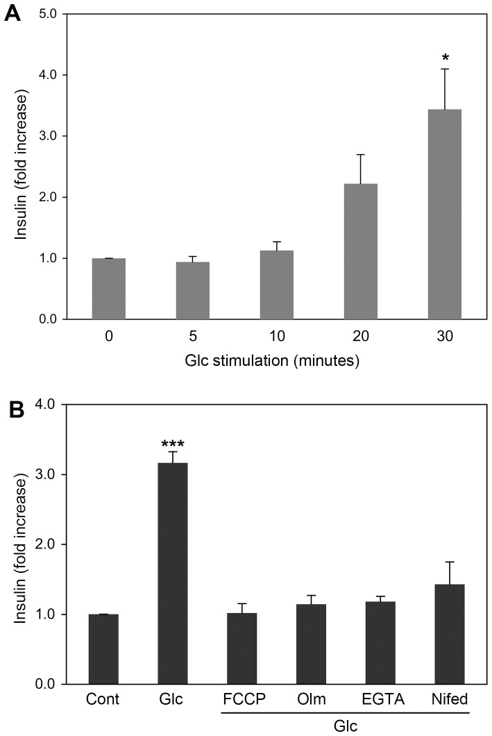
Figure 1. Glucose-stimulated insulin secretion requires mitochondrial function and Ca2+ influx.
(A) Insulin secretion of INS-1E cells was measured at different times of 20 mM glucose stimulation. Secreted insulin levels were determined by ELISA. Significant increases of insulin were detected in 30-minute glucose stimulation. (B) Insulin secretion was assayed in INS-1E cells incubated in 20 mM glucose for 30 min in the presence of FCCP, oligomycin (Olm), EGTA, or Nifedipine. Error bars represent SEM. n = 3. * and *** are for P<0.05 and P<0.001, respectively, with no glucose control.

