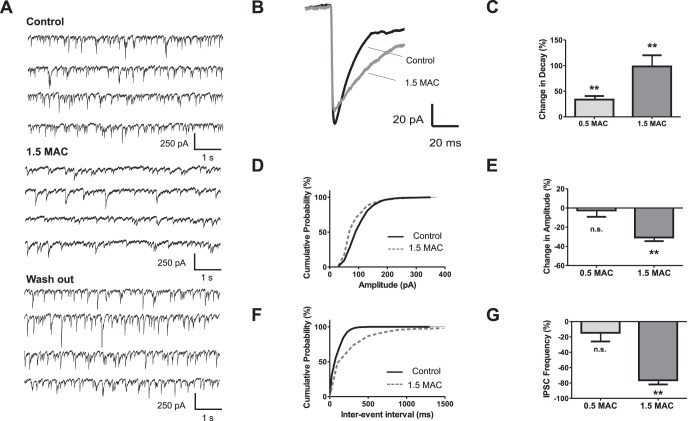
Figure 2. Frequency and amplitude of GABAA IPSCs is significantly reduced by high sevoflurane concentration.
(A) Sevoflurane effects on GABAA inhibitory postsynaptic currents (IPSCs). Original recording showing control condition (upper trace), sevoflurane (1.5 MAC, middle trace) and wash out (lower trace). GABAA IPSCs were isolated by adding CNQX (50 µM), AP5 (50 µM), and strychnine (1 µM). (B) Overlay of averaged IPSCs from the same experiments as depicted in (A). Note the prolonged decay time and the slightly reduced amplitude under sevoflurane (1.5 MAC). (C) In summary, application of sevoflurane significantly increased the decay times by 33.7±7.1% at 0.5 MAC (n = 8, p<0.01), and 98.4±21.8% at 1.5 MAC sevoflurane (n = 9, p<0.01). Absolute decay time values were 22.3±1.3 ms for control (n = 17), 32.2±2.5 ms for 0.5 MAC (n = 8), and 40.4±5.4 ms for 1.5 MAC (n = 9). (D) Cumulative distribution of IPSC amplitudes from the presented experiment. Sevoflurane shifted the population to smaller IPSC amplitudes (dotted gray line) compared with values from control condition (black line) (E) Pooled date showed that amplitudes of GABAA IPSCs remained stable at 0.5 MAC (change in amplitude −2.5±6.7%, n = 8), but were significantly changed by −30.7±3.8% at 1.5 MAC sevoflurane concentration (n = 9, p<0.01). (F) Cumulative distribution of inter-event intervals for shown representative traces. Sevoflurane prolonged the inter-event intervals (control black line, sevoflurane dotted gray line). (G) Analysis revealed that the IPSC frequency was essentially reduced at 1.5 MAC sevoflurane (change in frequency by −76.4±5.5% for 1.5 MAC, n = 9, p<0.01), whereas such an impressive effect was not detectable at 0.5 MAC sevoflurane (change in frequency −14.6±11.2%, n = 8). **p<0.01 as indicated.

