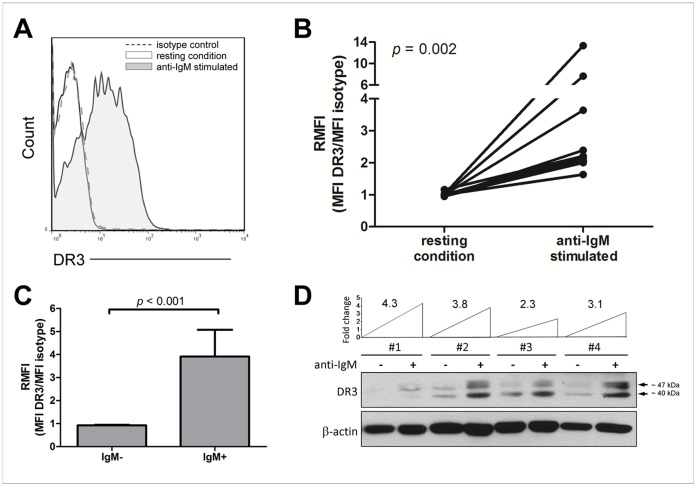
Figure 1. DR3 surface expression in B cells.
(A) Representative flow cytometry histograms of surface DR3 expression in purified B cells, in resting conditions or following stimulation with anti-IgM (n = 10). Analyses were gated on lymphocytes (based on forward and side scatter), living cells (7AAD-negative), and B cells (CD19-positive). (B) Surface DR3 expression in resting and BCR-stimulated B cells (n = 10). Data are expressed as DR3-expression median fluorescence intensity (MFI) divided by isotype-matched control (relative median fluorescence intensity = RMFI). Comparison between resting and anti-IgM-stimulated B cells was performed by the two-sample Wilcoxon signed rank sum test. (C) Surface DR3 expression in IgM-negative (IgM-) and IgM-positive (IgM+) B cells (n = 10). Data are expressed as difference in DR3-expression median fluorescence intensity (MFI) divided by isotype-matched control (relative median fluorescence intensity = RMFI). Comparison between IgM-negative and IgM-positive B cells was performed by the Mann Whitney test. Data are represented as mean ± SEM. (D) Western blot analysis of cell lysates of purified B cells (n = 4), in resting conditions or following stimulation with anti-IgM. The level of DR3 induction after anti-IgM stimulation is reported as fold change.

