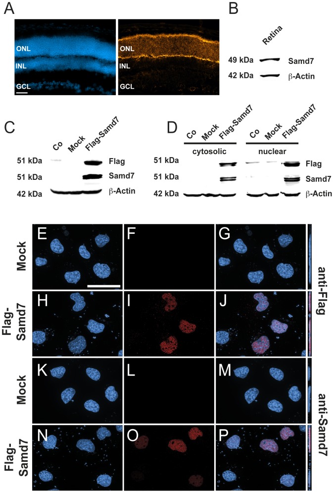
Figure 3. Samd7 is expressed in the outer nuclear layer of the mouse retina and localizes to the nucleus of transfected cells.
A: Immunhohistochemical analysis shows that Samd7 localizes to the outer nuclear layer in the adult mouse retina. Left panel: DAPI staining, right panel: anti-Samd7 antibody staining, ONL: outer nuclear layer, INL: inner nuclear layer, GCL: ganglion cell layer. Scale bar, 50 µm. B: Western blot performed with retinal lysates detecting Samd7 at a molecular weight of 49 kDa and beta-actin as loading control. C: Western blot performed with protein lysates from naive HEK293 cells (Co) or HEK293 cells transfected with mock plasmid or Flag-Samd7 expression plasmid. Anti-Samd7 antibody, anti-flag antibody, and anti-beta-actin antibody were used. The Flag-Samd7 band had a molecular weight of approximately 51 kDa. D: Western blot of cytosolic and nuclear fractions of HEK293 cells transfected with mock plasmid or Flag-Samd7 expression vector. Samd7 was was detected in both the cytosolic and nuclear fractions at a molecular weight of 51 kDa. (E–P): Subcellular localization of Samd7 in HEK293 cells transfected with Flag-Samd7 expression vector shown in fluorescent Z-stacked optical images. Mock transfected cells did not show a specific red signal with either the anti-Flag (F) or the anti-Samd7 (L) antibody. The anti-Flag antibody (I, J) as well as the anti-Samd7 antibody (O, P) showed a specific nuclear staining in Flag-Samd7 transfected cells when couter-stained with DAPI.

