
Fig. 1.
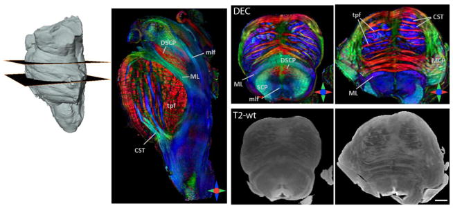
DTI of the ex vivo brainstem at 255 μm isotropic resolution. Sagittal and axial sections from the direction-encoded colormap (DEC) derived from DTI are shown. Corresponding T2-weighted (T2-wt) axial sections are shown for anatomic comparison. In the DEC maps, red, blue and green represent anisotropy along medial-lateral, superior-inferior, and anterior-posterior orientations, respectively. Abbreviations used are: CST: corticospinal tract, ML: medial lemniscus, mlf: medial longitudinal fasciculus, MCP: middle cerebellar peduncle, SCP: superior cerebellar peduncle, DSCP: decussation of the superior cerebellar peduncle, tpf: transverse pontine fibers. Scale bar = 4 mm.