
Fig. 3.
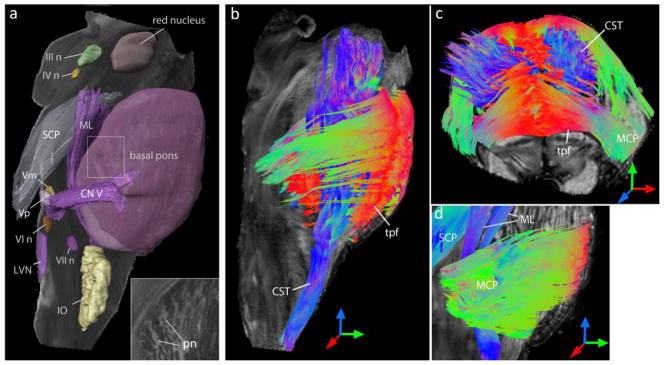
Three-dimensional reconstruction of brainstem nuclei and white matter tracts from the DTI data. a) Reconstructed cranial nerve and inferior olivary nuclei are overlaid on the ADC map for anatomic reference. The pontine nuclei in the basal pons are visible as hyperintense bands in the ADC map (inset). b–d) Three-dimensional rendering of fibers reconstructed from tractography are overlaid on fractional anisotropy images for anatomic reference, with color representing the orientations of the fibers. Red, green and blue indicate fiber orientations along the medial-lateral, anterior-posterior, and superior-inferior axes, respectively. Abbreviations are: III n: oculomotor nucleus, IV n: trochlear nucleus, Vp: primary sensory nucleus of trigeminal nerve, Vm: trigeminal motor nucleus, VI n: abducens nucleus, VII n: facial motor nucleus, CN V: trigeminal nerve, CST: corticospinal tract, IO: inferior olivary nucleus, LVN: lateral vestibular nucleus, MCP: middle cerebellar peduncle, ML: medial lemniscus, pn: pontine nuclei, SCP: superior cerebellar peduncle, tpf: transverse pontine fibers.