
Fig. 5.
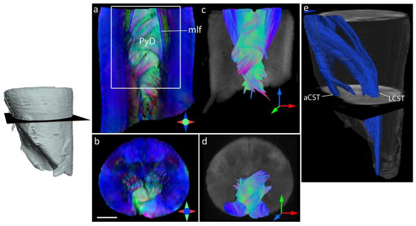
Ex vivo DTI of the cervical spinal cord at the level of the spinomedullary junction showing the decussation of the pyramidal tracts (PyD). a–b) Coronal and axial sections through the DEC maps show delineation of the PyD and medial longitudinal fasciculus (mlf). Scale bar = 2 mm. c–d) Fiber tractography shows interdigitating bundles from the left and right pyramidal (Py) tracts (reconstructed fibers are cropped to coronal and axial sections in c and d, respectively). Fibers are colored by orientation, with red, green and blue denoting fiber orientations along medial-lateral, anterior-posterior, and superior-inferior axes, respectively. e) Three-dimensional reconstruction of the left pyramidal tract (rendered in blue), showing crossed fibers forming the lateral corticospinal tract (LCST) and uncrossed pyramidal fibers continuing into the ipsilateral anterior corticospinal tract (aCST).