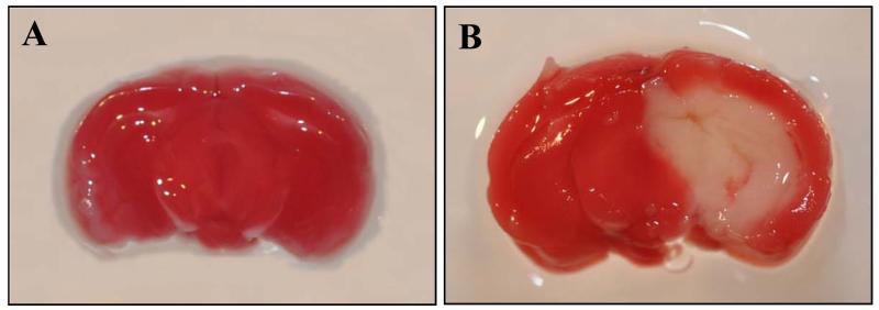
Fig. 1.
Induction of brain injury in the mouse after MCAO. Coronal slices of fresh brain tissue were stained with triphenyl tetrazolium chloride. Infarct area of the brain appears as pale staining on the slice and viable brain area shows plum red color. A representative brain slice from (A) sham-operated animals and (B) 24 h after subjecting to MCAO.

