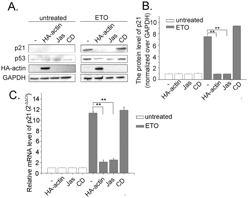
Figure 6. The impact of actin polymerization on p53 leads to the alteration of p21 expression.
A. Cells were transfected with HA-actin or treated with Jas (50 nM) or CD (0.01 µg/ml) for 2 h. Then, cells were treated with ETO (10 µM) or untreated as control for 12 h. Whole cell proteins were extracted and western blotting was performed to measure p21 protein levels. B. Western blotting were analyzed with Image J software, and the results are presented as mean ± SD of values from three independent experiments. C. Cells were transfected with HA-actin or treated with Jas (50 nM) or CD (0.01 µg/ml) for 2 h. Then, cells were treated with ETO (10 µM) or untreated as control for 12 h. Real-Time PCR was performed, mRNA content of p21 was normalized to that of GAPDH and the normal cells’ mRNA level was valued as 1. Data (mean±SD) were from three independent experiments. All Statistical differences were determined by One-way ANOVA. **, P<0.01.

