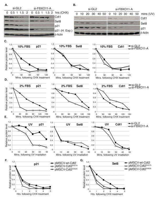
Figure 5. Inactivation of FBXO11-mediated degradation of Cdt2 increases the turnover of the Cdt2 substrates p21 and Set8.
(A–E) Measurement of the half-life (t1/2) of various CRL4Cdt2 substrate proteins following the depletion of FBXO11 from asynchronously proliferating U2OS cells cultured in 10% (A, C) or 2% serum (D) or in U2OS cells cultured in 10% serum and irradiated with 30J/m2 UV (B, E).
(A, B) Immunoblot of Cdt1, Set8 and p21 in extracts of U2OS cells cultured in 10% serum and transfected with indicated siRNAs following CHX addition for the indicated hours (A), or at the indicated time following UV irradiation (B). β-actin: loading control.
(C–E) t1/2 of p21, Set8 or Cdt1 in cells grown in 10% serum (C), 2% serum (D) or following UV irradiation (E). (C and E) Quantitation of indicated proteins shown in (A and B, respectively), normalized to β-actin and expressed relative to the 0 time point in each case. (D) Same as (C), except U2OS cells were cultured in 2% serum for 24 hr prior to the CHX addition. Western blots not shown. See also Figure S3.
(F, G) FBXO11-resistant Cdt2 proteins destabilize p21 and Set8. Quantitation of p21 (F) and Set8 (G) proteins in extracts of U2OS cells stably expressing wt-Cdt2, Cdt2D457A or Cdt2S462A from pMSCV-based retrovirus vectors. Values are normalized to those of β-actin and expressed relative to the 0 time point when CHX is added.

