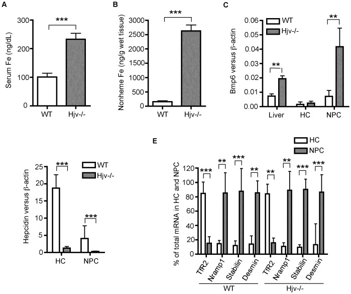
Figure 1. Increased Bmp6 mRNA expression in the liver of Hjv-/- mice is mainly detected in the non-parenchymal cells.
Five male wild type (WT) and five Hjv-/- mice at ∼10-weeks old were used for the studies. A. Serum iron. Serum iron concentrations were measured using a serum iron/TIBC Reagent Set (Teco Diagnostics, Anaheim, CA) according to the manufacturer's instructions. Each sample was measured twice in triplicate. Serum iron concentrations are expressed as microgram per deciliter (dL). B. Liver nonheme iron. Nonheme iron concentrations in the liver tissues were measured after digestion with acid buffer. Each sample was measured twice in triplicate. Iron concentrations are expressed as microgram per gram wet tissue. C. qRT-PCR analysis of Bmp6 mRNA in the whole liver tissues, isolated hepatocytes (HC), and the total non-parenchymal liver cells (NPC) from WT and Hjv-/- mice. The results are expressed as the amount of mRNA relative to β-actin in each sample. D. qRT-PCR analysis of hepcidin mRNA in the whole liver tissues, isolated hepatocytes (HC), and total NPC from WT and Hjv-/- mice. The results are expressed as the amount of mRNA relative to β-actin in each sample. E. qRT-PCR analysis of Tfr2 (a specific marker for hepatocytes), Nramp1 (a specific marker for KCs), stabilin-1 (Stabilin, a specific marker for SECs), and desmin (a specific marker for HSCs) mRNA in the isolated hepatocytes (HC) and total NPC from WT and Hjv-/- mice. The mRNA levels were first calculated as the amount relative to β-actin in each sample. For each gene of interest, the expression levels in hepatocytes (HC) and NPC were then converted to percentages using the total amount in both HC and NPC as a whole and presented. **, P<0.01; ***, P<0.001.

