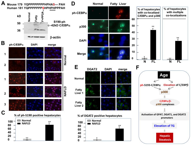
Figure 7.
C/EBPα-p300-DGAT1/2 pathway is elevated in livers of patients with non-alcoholic fatty liver disease. A. Generation of antibodies to human p-Ser190 isoform of C/EBPα. Sequences of mouse and human C/EBPα surrounding Ser193 and Ser190 are shown. Asterisks show differences in the sequences. Bottom: Western blotting with antibodies to p-Ser190 human C/EBPα. F+PP2A; proteins from patients with fatty liver were treated with phosphatase PP2A. B. Immunostaining of normal livers and livers from patients with fatty liver diseases using antibodies to p-Ser190 isoform of C/EBPα. The slides were stained with DAPI. C. Percentage of p-Ser190-C/EBPα positive hepatocytes in livers of normal patients and in livers of patients with fatty liver disease. D. Amounts of p-Ser190–C/EBPα-p300 complexes are increased in livers of NAFLD patients. The same liver sections were stained with antibodies to p-Ser190-C/EBPα, to p300, and with DAPI. Merge shows colocalization of C/EBPα and p300 in nucleus. Bar graphs show total number of hepatocytes with colocalization of C/EBPα and p300 and number of hepatocytes with multiple foci of colocalizations. E. Amounts of DGAT2 are increased in livers of patients with fatty liver diseases. Liver sections were stained with antibodies to DGAT2 and with DAPI. Bar graphs show percentage of DGAT2-positive hepatocytes. F. A model showing hypothetical mechanisms for the age-associated hepatic steatosis.

