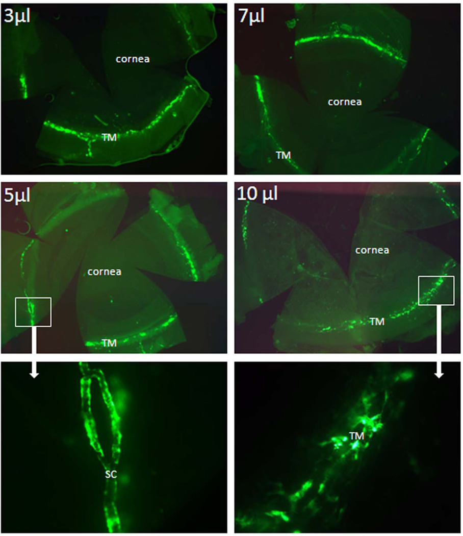
Figure 3. GFP expression in mouse anterior segments following rapid intracameral infusion of adenovirus encoding GFP.
Ad.cmv.gfp (5×10⁵ particles in 3, 5, 7, or 10 µl volume) was infused into mouse anterior chambers over 3 minutes (flow rate 1, 1.7, 2.34 or 3.33 µl/min). Three to five days after transduction, mouse eyes were fixed and dissected. The anterior segments were flat mounted and the images were captured with a fluorescence microscope. Note: top four panels (2.5 × amplification); bottom two panels (20 × amplification). The images are representative results of three to five individual experiments for each condition using both CD1 and C57BL/6 mice (8–30 weeks old).

