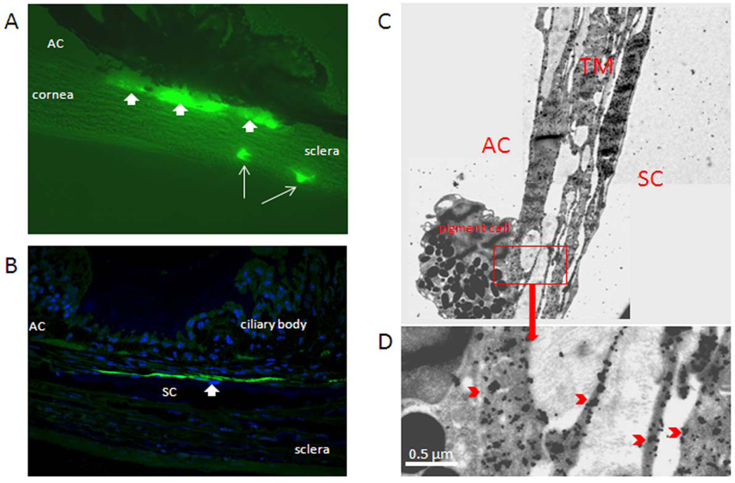
Figure 4. GFP expression in mouse aqueous humor outflow pathway following fast intracameral infusion of adenovirus encoding GFP.
Ad.cmv.gfp (5×10⁵ particles in 10 µl volume) was infused into mouse anterior chamber over 3 minutes (flow rate 3.33 µl/min). Three to five days after transduction, mouse eyes were fixed and dissected. The anterior segments were then processed by cryosectioning (10 µm), semi-thin sectioning (0.5 µm) or thin sectioning (65 nm). A) Immunofluorescence image of sagittal plane section through mouse angle structures; B) Sagittal plane semi-thin section (60 × amplification) through angle structures of mouse eye. The section was immunostained with anti-GFP primary antibody and Alexa Fluor®488 goat anti-rabbit IgG (H+L) secondary antibody. Nuclei were stained with DAPI. The image was captured with a confocal microscope; C) Sagittal plane thin section through trabecular meshwork and Schlemm's canal. The section was immunostained with specific anti-GFP primary antibodies and 6 µm immunogold anti-rabbit secondary antibodies. Images were captured with a JEM-1400 electron microscope. Data are representative of 3 experiments using C57BL/6 mice (8–22 weeks old).

