Figure 5.

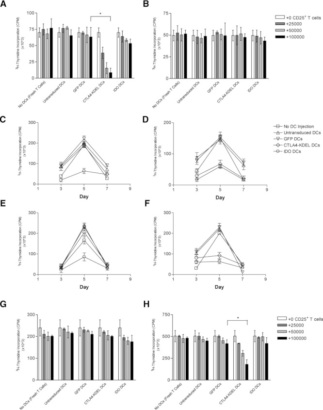
Generation of Treg cells in vivo with indirect donor allospecificity and capacity for linked suppression. 2.5 × 10⁶ CBK DCs (either untransduced, or transduced with EIAV-GFP (control), EIAV-CTLA4-KDEL or EIAV-IDO) were injected i.v. into C3H/He mice. (A–B) After 10 days, CD4+CD25+ T cells purified from the spleens were irradiated and added (0–105 CD4+CD25+ T cells) to a primary MLR between freshly isolated CBA/Ca-derived CD4+ T cells and (A) donor CBK DCs or (B) third-party B10.A DCs. T-cell proliferation was assessed by thymidine incorporation after 5 days. (C–D) CD4+ T cells purified from the spleens of injected mice were also rechallenged in vitro with (C) (BALB/c × C57BL/6)F1 DCs expressing only third-party MHC or (D) (C57BL/6 × CBA/Ca)F1 DCs expressing both donor (Kb presented in the context of I-Ak/I-Ek) and third-party MHC, and CD4+ T-cell proliferation was assessed by thymidine incorporation on days 3, 5 and 7. (E–F) Rechallenge assays were repeated using CD4+ T cells that were depleted of CD4+CD25+ T cells prior to rechallenge with (E) (BALB/c × C57BL/6)F1 DCs or (F) (C57BL/6 × CBA/Ca)F1 DCs. (G–H) In addition, CD4+CD25+ T cells purified from the spleens of injected mice were irradiated and added (0–105 CD4+CD25+ T cells) to new primary MLRs between freshly isolated CBA/Ca-derived CD4+ T cells and (G) (BALB/c × C57BL/6)F1 DCs or (H) (C57BL/6 × CBA/Ca)F1 DCs and T-cell proliferation assessed by thymidine incorporation after 5 days. All results are shown as the mean ± SD of triplicate wells and are representative of three independent experiments performed. *p < 0.05, two-tailed t-test.
