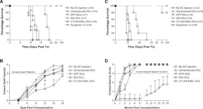
Figure 7.

Prolongation of corneal graft survival following administration of modified DCs. BALB/c mice were either untreated (n = 6) or given 2.5 × 10⁶ C3H/He DCs intravenously. The DCs were either untransduced (n = 5), or transduced 72 h earlier ex vivo with equine infectious anaemia virus (EIAV)-GFP (control, n = 7), EIAV-IDO (n = 7) or EIAV-CTLA4-KDEL (n = 8). Ten days later, the BALB/c mice received a complete MHC-disparate C3H/He corneal graft or a syngeneic BALB/c graft (n = 6). (A) Data were plotted using the Kaplan–Meyer method and differences in graft survival were analysed using a log-rank test. (B) Corneal graft opacity scores were plotted and statistical differences between CTLA4-KDEL- and GFP (control)-expressing DCs calculated using the Mann–Whitney U test. * p < 0.008. (C) Using the same protocol, BALB/c mice were either untreated (n = 4) or given 2.5 × 10⁶ (CBA × BALB/c)F1 DCs intravenously. The DCs were either untransduced (n = 5), or transduced 72 h earlier ex vivo with EIAV-GFP (n = 5), EIAV-IDO (n = 5) or EIAV-CTLA4-KDEL (n = 5). 10 days later, BALB/c mice received a complete MHC-disparate CBA/Ca corneal graft or a syngeneic BALB/c graft (n = 6). Data were analysed as above. (D) Corneal graft opacity scores were plotted and statistical differences between CTLA4-KDEL- or IDO-expressing DCs and GFP-expressing DCs calculated using the Mann–Whitney U Test. *p < 0.008. Data shown are representative of three independent experiments performed.
