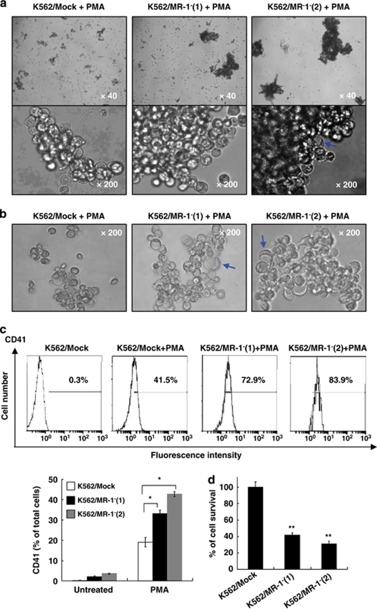
Figure 4.
MR-1 silencing promoted MK differentiation induced by PMA. K562/MR-1− cells were treated with 20 nℳ PMA for 48 h (a) or for 96 h (b), and then the megakaryocytic features were analyzed by light microscopy. After 96-h treatment, CD41 expression (c) and cell viability (d) were measured by FACS and cell count assay, respectively. *P<0.05; **P<0.01.

