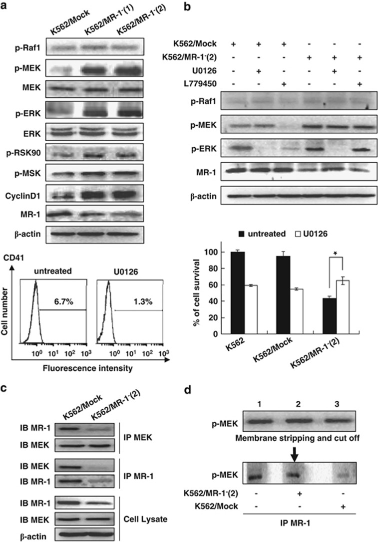
Figure 5.
The MEK/ERK signal involved in MK differentiation by MR-1-mediated MEK dephosphorylation. Western blot analysis of various signal proteins. (a) Cells were treated with 20 μℳ U0126 for 2 h or with 10 μℳ L779450 for 1 h. Various signal proteins were detected by WB (up panel), and CD41 was analyzed by FACS; cell viability was monitored by cell count (b). Interaction of MR-1 and MEK was analyzed by reciprocal immunoprecipitation (c). Triplicate cell lysates from K562/Mock were subjected to western blot for p-MEK detection, then stripped and cut into thirds, each lane containing an equal amount of p-MEK incubated with IP MR-1 from blank control (lane 1), Mock/K562 (lane 2) or K562/MR-1− cell lysate (lane 3) for 30 min, followed by p-MEK western blot analysis. (d) The data are the means±s.d. of two experiments; significance of Student's t-test: *P<0.05.

