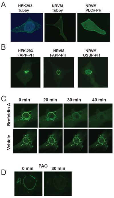
Figure 6.
PI4P localizes to perinuclear Golgi surrounding the nuclear envelope in cardiac myocytes. (A) Detection of PI4,5P2 localization in NRVMs. Left panel: HEK-293 cells transfected with Tubby GFP and stained with DAPI, middle: NRVMs transfected with Tubby-GFP and right: NRVMs transfected with PLCδ-PH-GFP and analyzed by confocal microscopy. (B) Detection of PI4P at the nuclear envelope. The indicated cell types were transfected with either OSBP-PH-GFP or FAPP-PH-GFP and analyzed by confocal microscopy. (C) Inhibition of ARF eliminates perinuclear staining with FAPP-PH-GFP. NRVMs transfected with FAPP-PH-GFP were treated with 100 ng/mL Brefeldin A and GFP fluorescence monitored with time. Cells treated with vehicle are shown in the bottom panels indicate a lack of photobleaching in these experiments. (D) Inhibition of PI4kinase with PAO depletes perinuclear fluorescence associated with FAPP-PH-GFP. NRVMs transfected with FAPP-PH-GFP were treated with 10 μM PAO and fluorescence analyzed by confocal microscopy. All experiments were repeated a minimum of 3 times. See also Figure S4.

