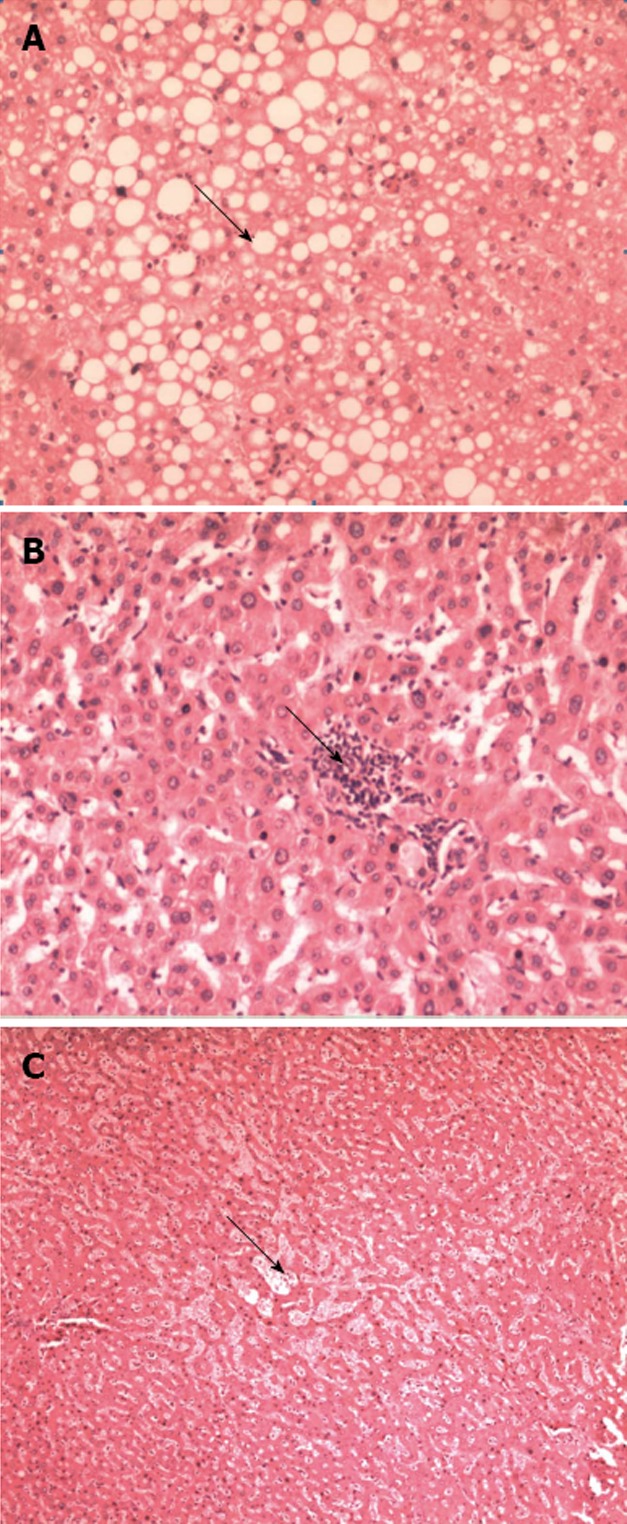
Figure 1.

Histopathological findings. A: Severe steatosis. Large drop of fat (arrow) in the majority of hepatocytes, HE, 100 ×; B: Example of steatohepatitis showing the foci (arrow) of inflammation among the hepatocytes, HE, 100 ×; C: Grade 3 sinusoidal dilation involved the complete lobule, HE, 40 ×.
