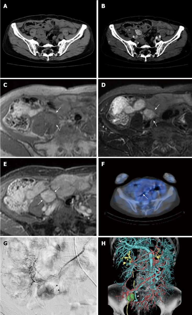
Figure 1.

Imaging features of tumor (white arrows) before treatment. A: Axial plain; B: contrast-enhanced computed tomography (CT), CT shows a smoothly marginated, heterogeneously enhanced tumor adjacent to the right major psoas muscle, 16 mm × 22 mm × 25 mm in size; C: T1-weighted magnetic resonance image shows a well defined, isointense mass; D: On T2-weighted images, the mass shows heterogeneous high intensity; E: On T1-weighted images after a bolus infusion of gadolinium chelate, the mass had marked contrast enhancement; F: Positron emission tomography-CT scan was negative; G: Superior mesenteric arteriography displays a markedly hypervascular mass (black arrow heads) adjacent to the terminal ileum; H: Volume rendering image acquired from angio-CT (white arrow heads).
