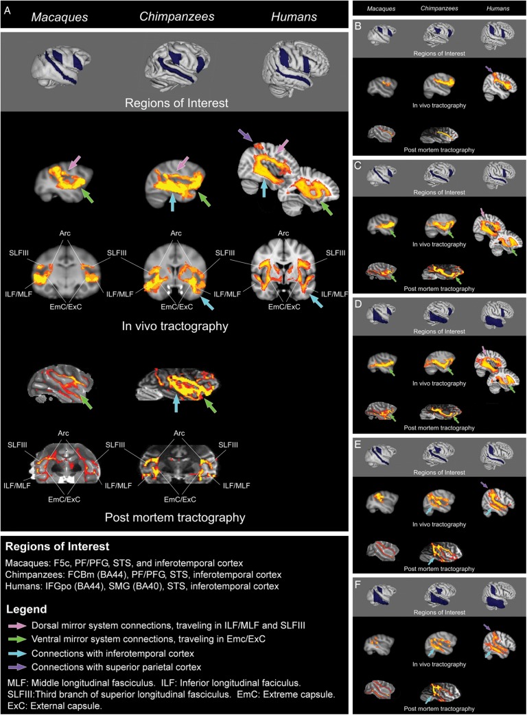
Figure 1.
(A) The mirror system as a whole: Connections between frontal mirror region, parietal mirror region, and superior temporal sulcus. Probabilistic tractography between macaque F5, PF, and superior temporal sulcus; chimpanzee BA44, PF, and superior temporal sulcus; and human BA44, BA40, and superior temporal sulcus. Three major species differences are apparent. First, there is an increase from macaques to chimpanzees to humans in the ratio of dorsal versus ventral connections within the circuit (inferior/middle longitudinal fasciculi (ILF/MLF) and third branch of superior longitudinal fasciculus (SLFIII) versus extreme and external capsules (EmC/ExC); pink versus green arrows). Second, in humans and chimpanzees, but not in macaques, this circuit includes a robust connection to inferior and middle temporal areas associated with object and tool recognition (blue arrows). Third, in humans only, this circuit includes a connection to superior parietal cortex, a region associated with spatial attention and tool use (purple arrow). Images are shown in radiological convention (right side of image corresponds to left side of brain). See online version for color figures. (B) Connections between frontal and parietal mirror regions. Probabilistic tractography between macaque F5 and PF, chimpanzee BA44 and PF, and human BA44 and BA40. These connections follow the third branch of the superior longitudinal fasciculus in all 3 species. In humans, this tract appears more robust, and includes a connection with superior parietal cortex (purple arrow). (C) Connections between superior temporal sulcus and frontal mirror region. Probabilistic tractography between macaque F5 and superior temporal sulcus; chimpanzee BA44 and superior temporal sulcus; and human BA44 and superior temporal sulcus. In all 3 species, connections between these regions follow the extreme/external capsules and pass through more anterior regions of prefrontal cortex en route to the frontal mirror region (green arrows). In humans, a second, dorsal pathway is detected, which travels through the inferior/middle longitudinal fasciculi through inferior parietal cortex to the third branch of the superior longitudinal fasciculus (pink arrow). Connections through the arcuate fasciculus (not shown here) were also detected, but this tract travels deeper in the white matter beneath parietal cortex and does not reach parietal gray matter. (D) Connections between inferior temporal cortex and frontal mirror region. Probabilistic tractography between macaque F5 and inferior temporal cortex ROI (inferior temporal and fusiform gyri); chimpanzee BA44 and inferior temporal ROI (middle, inferior, and fusiform gyri); and human BA44 and inferior temporal ROI (middle, inferior, and fusiform gyri). In all 3 species, connections between these regions follow the extreme/external capsules and pass through more anterior regions of prefrontal cortex en route to the frontal mirror region (green arrows). In humans, a second, dorsal pathway is detected, which travels through the inferior/middle longitudinal fasciculi through inferior parietal cortex to the third branch of the superior longitudinal fasciculus (pink arrow). Connections through the arcuate fasciculus (not shown here) were also detected, but this tract travels beneath the white matter of parietal cortex and does not reach parietal gray matter. (E) Sub-connections between superior temporal sulcus and parietal mirror region. Probabilistic tractography between macaque PF and superior temporal sulcus; chimpanzee PF and superior temporal sulcus; and human BA40 and superior temporal sulcus. In all 3 species, these connections follow the middle longitudinal fasciculus, but these connections extend further in chimpanzees than macaques, and furthest in humans. Connections with inferior temporal cortex via the inferior/middle longitudinal fasciculi are also apparent in chimpanzees and humans, and are more robust in humans (blue arrows). These connections also included a projection to superior parietal cortex in humans (purple arrow). (F) Connections between inferior temporal cortex and parietal mirror region. Probabilistic tractography between macaque PF and inferior temporal ROI (inferior and fusiform gyri); chimpanzee PF and inferior temporal ROI (middle, inferior, and fusiform gyri); and human BA40 and inferior temporal ROI (middle, inferior, and fusiform gyri). These connections travel through the inferior/middle longitudinal fasciculi in all 3 species. They are quite weak in macaques, robust in chimpanzees, and most robust in humans (blue arrows). In humans, a connection with superior parietal cortex is also apparent (purple arrow).

