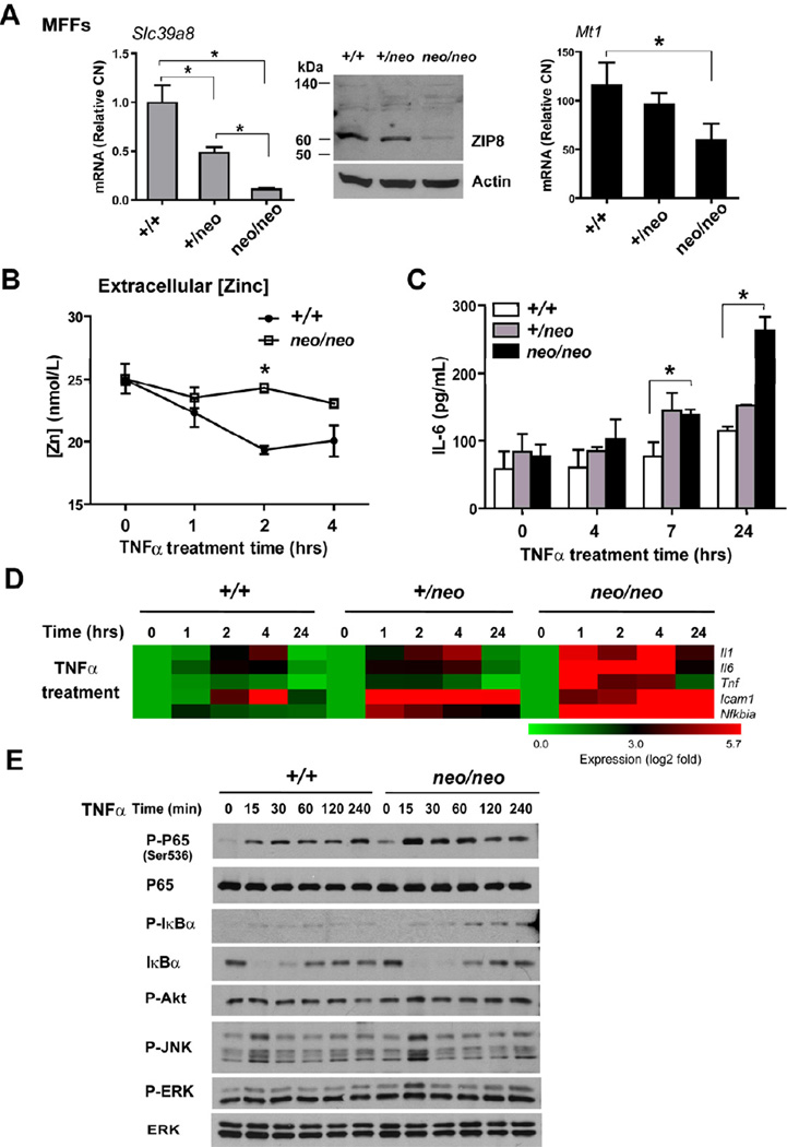
Figure 7. Slc39a8 hypomorphic mouse fetal fibroblasts (MFFs) have an elevated pro-inflammatory response to TNFα.
(A) Real-time PCR quantified the mRNA levels of ZIP8 and MT1 in Slc39a8 (+/+), Slc39a8 (+/neo) and Slc39a8 (neo/neo) primary MFFs cultures. Western analysis shows ZIP8 protein levels. (B) Total zinc levels in culture medium were determined by AAS. (C) Slc39a8 (+/+), Slc39a8 (+/neo) and Slc39a8 (neo/neo) MFFs were treated with mouse TNFα (50 ng/mL) for the indicated time. IL-6 levels were measured in the culture supernatants. (D) Analysis of pro-inflammatory gene expression profiles in Slc39a8 (+/+), Slc39a8 (+/neo) and Slc39a8 (neo/neo) MFFs that were treated with TNFα for the indicated time points. Data are presented as a representative heatmap following normalization to untreated group. Gene designation is to the right of each row. (E) Western analysis of signaling pathways in Slc39a8 (+/+) and Slc39a8 (neo/neo) MFFs treated with TNFα.

