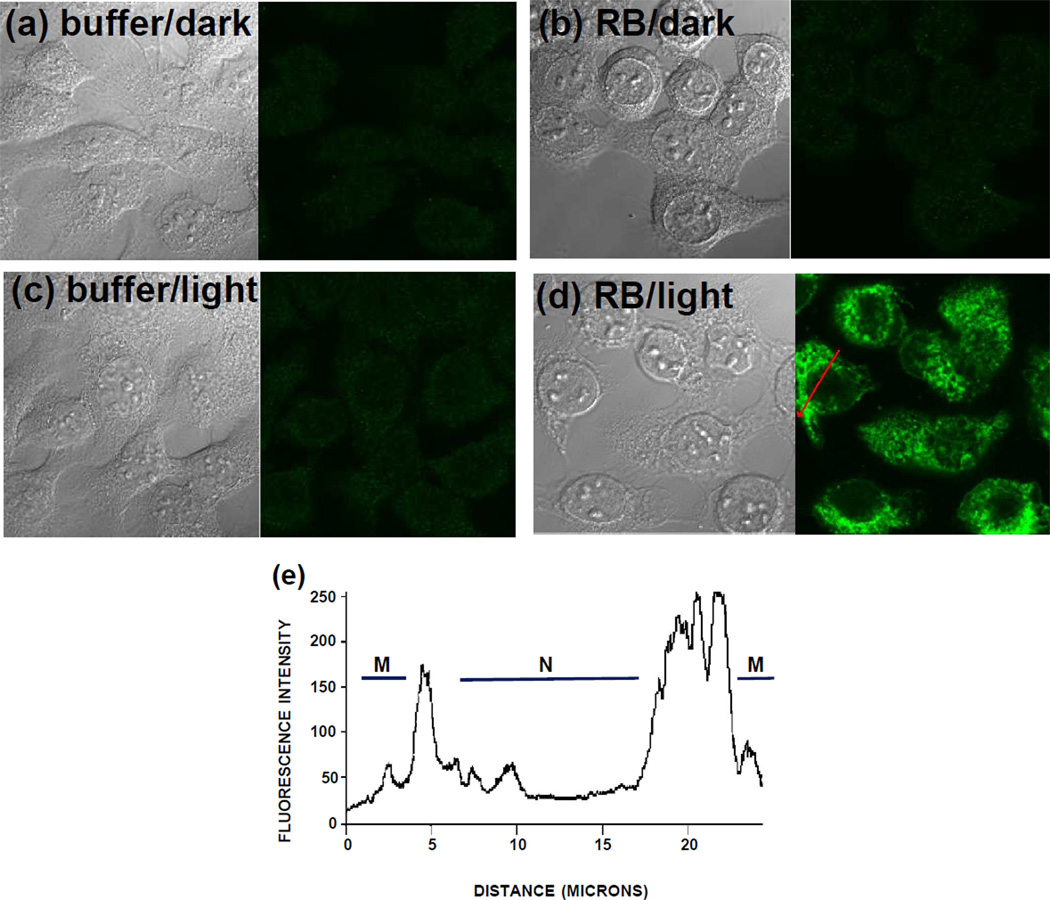
Figure 2.
Confocal images of HaCaT cells stained with anti-NFK antibody and Alexa 488 secondary antibody and visualized using a 63X oil immersion lens. Cells incubated with 10 µM rose bengal (RB) (b, d) or buffer (a, c) for 1 hour in the dark were washed free of the dye and were then exposed to 20 min incubation under cool white fluorescent lights (c, d) or were maintained in the dark (a, b). Paired images show transmission microscopy on the left side and confocal fluorescence microscopy on the right side. (e) Pixel intensity across a cell in (d) as indicated by red arrow. M, membrane; N, nucleus.

