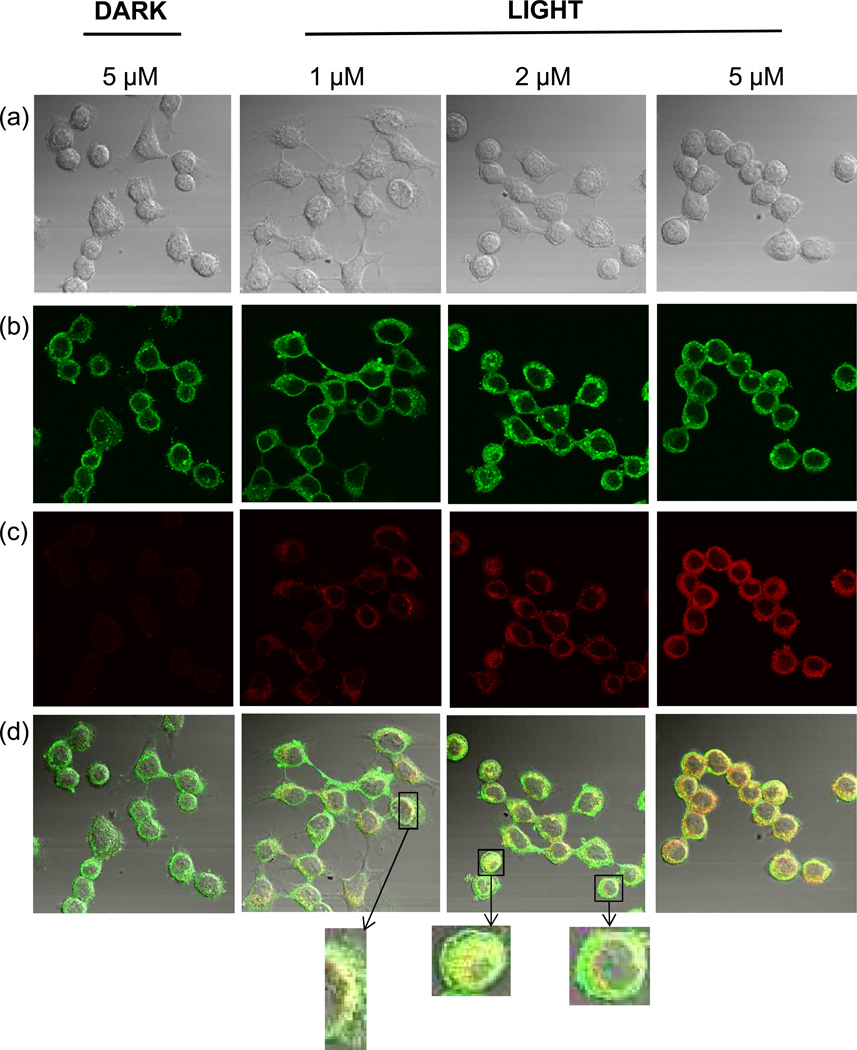
Figure 3.
Partial colocalization of NFK with Golgi in HaCaT cells after photosensitization with rose bengal. HaCaT cells were incubated with 1, 2, or 5 µM rose bengal as indicated. After washing, cells were exposed to 20 min incubation under cool white fluorescent lights or were maintained in the dark as indicated. Cells were stained with anti-NFK antibody and anti-Golgi-97 antibody followed by Alexa 488 and Alexa 568 antibody. (a) DIC image (transmission microscopy), (b) anti-golgin-97 (green), (c) anti-NFK (red), and (d) merge of (a), (b) and (c).

