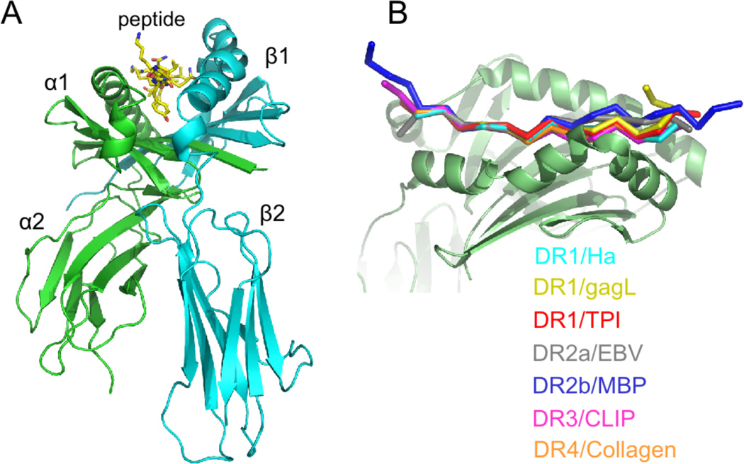
Figure 3.
Structure of the class II MHC – peptide complex. (A) Extracellular domain containing alpha (green) and beta (cyan) subunits and bound peptide. Short peptide sequences connecting the extracellular domain to the membrane spanning regions extend from the alpha and beta termini at the bottom of the figure. (B) Overlay of DR1-peptide complexes, with peptide N-terminus to the left and the MHC beta subunit in front. All peptides adopt essentially the same conformation, although variation at the peptide termini can be seen.

