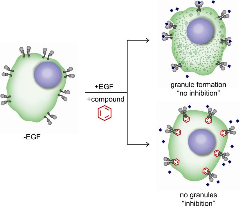
Figure 1. Principles of the EGFRB assay.
Schematics of the EGFRB assay with A549 EGFR biosensor cell line (A549-EGFRB). In absence of EGF stimulation, diffused GFP is observed in the cytoplasm of cells. In contrast, EGF addition triggers EGFR activation and subsequent clustering and internalization as observed by the formation of granules (vesicles) in the GFP channel, corresponding to “no inhibition”. Granule formation upon EGF stimulation is prevented by EGFR small molecule inhibitors (“inhibition”), allowing the identification of novel EGFR inhibitors by HTS.

