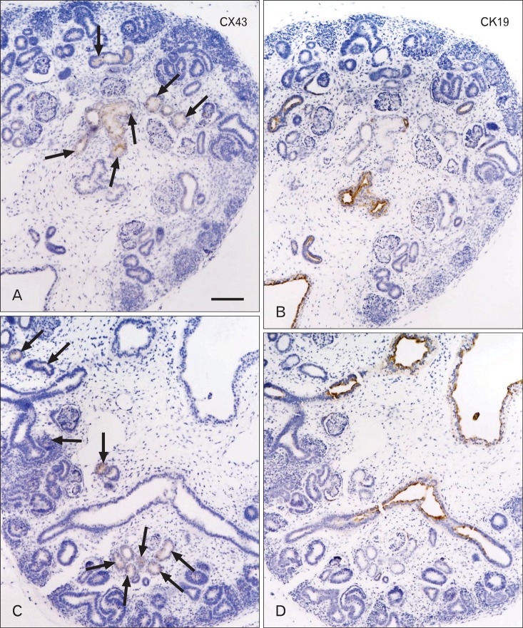
Fig. 3.
Immunohistochemical analysis for cytokeratin and connexin in the human fetal kidney: a fetus at 11 weeks. Panels (A) and (B) and panels (C) and (D) show adjacent sections. Connexin-43 (CX43) immunopositivity is seen in some of the renal tubules of the cortex (arrows in A and C), whereas cytokeratin-19 (CK19) shows strong immunoreactivity in the collecting ducts and renal pelvis (B, D). Scale bar in (A)=0.2 mm (A-D).

