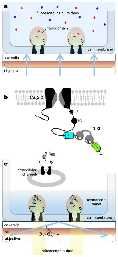
Figure 1. Approach to resolving channel nanodomain Ca2+ signals.
a, Conventional wide-field imaging using cytosolic chemical fluorescent dyes cannot resolve channel nanodomain Ca2+ signals. Blue shading denotes region of fluorescence excitation, which extends throughout the cell under wide-field imaging. b, Design of the genetically encoded Ca2+ indicator TN-XL fused to the carboxy tail of the α1B subunit of a CaV2.2 channel, yielding CaV2.2/TN-XL. For orientation, structure-function elements involved in calmodulin regulation are denoted on carboxy terminus21: EF, EF-hand region; IQ, IQ-domain for apoCaM binding. CFP denotes enhanced CFP. YFP denotes circularly permuted Citrine. c, CaV2.2/TN-XL constructs act as a ‘near-field’ sensor of nanodomain Ca2+. TIRF imaging evanescent wave illuminates only CaV2.2/TN-XL channels within ~150 nm from the glass/cell membrane interface, as indicated by the blue-shaded region. This mode of excitation potentially excludes intracellular channels from consideration. When laser illumination angle Θ exceeds a critical angle ΘC, TIRF illumination occurs.

