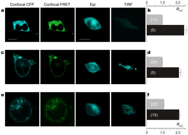
Figure 2. CaV2.2/TN-XL fusion construct preserves function of sensor.
a, Behavior of free TN-XL. Left to right: Confocal (CFP filter), confocal (FRET filter), epi-fluorescence (CFP filter), and TIRF (CFP filter) images of HEK293 cells expressing cytoplasmic TN-XL. White scale bar indicates 10 μM. Bar at far left pertains to all confocal images. Bar at middle right pertains to all epifluorescence and TIRF images. b, TN-XL ratio (RF/C=SF/SC) measured in resting cells (Rmin) (gray bars) and in cells under high Ca2+ (Rmax) (black bars). Data shown as mean ± sem with number of cells in parentheses. c and d, Behavior of TN-XL-Ras (membrane targeted TN-XL). Format as in a and b, respectively. e and f, Behavior of CaV2.2/TN-XL (N-type channel fused to TN-XL). Format as in a and b, respectively.

