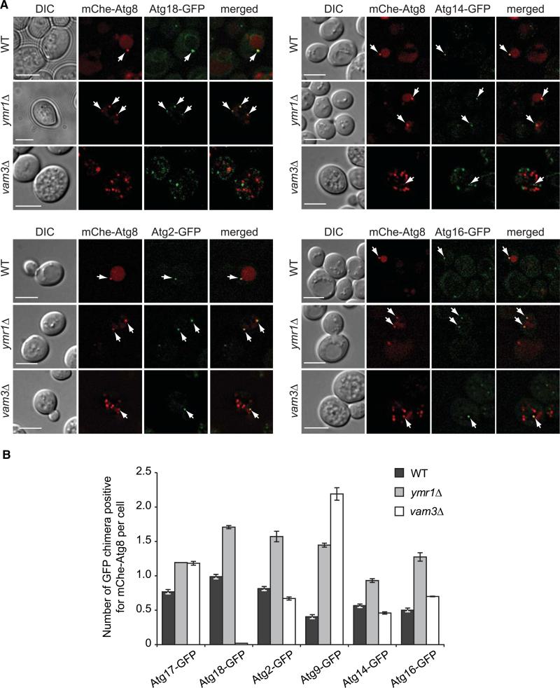
Figure 5. Atg Proteins Remain Associated with Autophagosomes in the ymr1Δ Strain.
WT, ymr1Δ, and vam3Δ strains expressing endogenous Atg2-GFP (PSY102, ECY155, and ECY183), Atg9-GFP (FRY162, ECY153, and AVY078) Atg14-GFP (PSY142, ECY162, and ECY184), Atg16-GFP (KTY148, ECY157, and ECY185), Atg17-GFP (ECY167, ECY169, and ECY172), or Atg18-GFP (PSY62, ECY137, and ECY147) and the pCumCheAtg8(415) plasmid were treated with rapamycin for 3 hr and imaged.
(A) Fluorescence microscopy images of the various strains. Arrows highlight colocalizations. DIC, differential interference contrast. Scale bars represent 5 μm.
(B) Quantification of the experiments presented in (A) and Figure S4A by determining the average number of GFP chimera positive for mCheAtg8 per cell. Error bars represent the SEM.
See also Figure S4.

