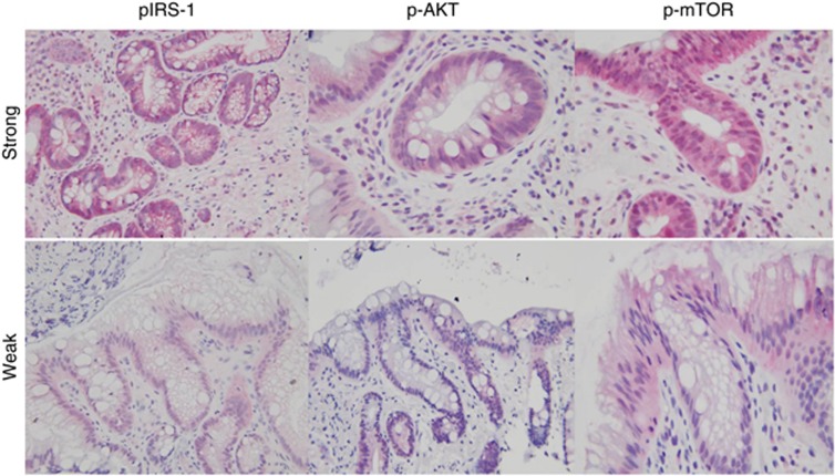
Figure 2.
Transmitted light micrographs of tissue derived from Barrett's esophagus (BE). Upper panel shows staining of tissues derived from BE, which were graded ‘strong' for the expression of the indicated antibody, indicating that >50% of the visualized field had positive immunoreactivity. The lower panel shows BE that was minimally reactive for the relevant antibody. AKT, protein kinase B; IRS, insulin receptor substrate; mTOR, mammalian target of rapamycin.

