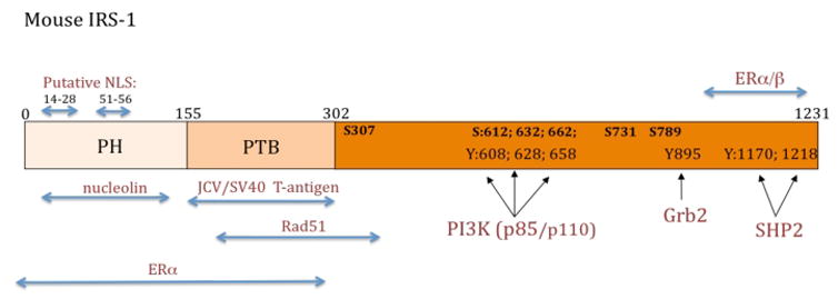
Figure 1. Schematic diagram of mouse IRS-1 protein.

There are two major functional domains within the N-terminus portion of IRS-1: pleckstrin homology domain (PH), spanning between amino acids 0–155, and phosphotyrozine binding domain (PTB) located between amino acids 155–302. Black arrows indicate exact binding sites for PI3 kinase, Grab2 and SHP2 at indicated tyrosine residues (Y). Functionally relevant serine residues (S) and their corresponding amino acid positions are also indicated. Blue arrows indicate putative binding regions for proteins, which are suspected to translocate IRS-1 to the nucleus, including nucleolin, polyomavirus T-antigens (JCV and SV40), estrogen receptor α (ERα) and estrogen receptor β (ERβ). Other indicated sites include the binding between IRS-1 and DNA repair protein, Rad51, and positions of putative nuclear localization signals (NLS).
