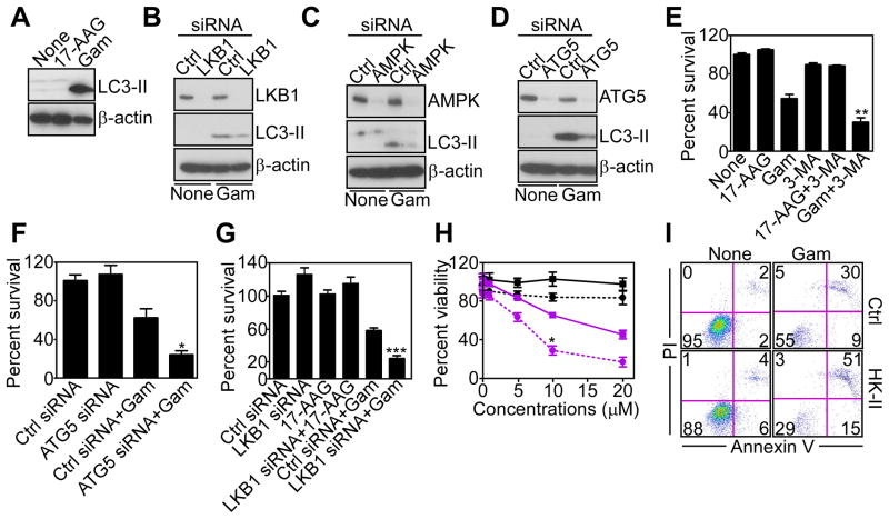
Figure 4. Mitochondrial proteotoxic stress stimulates autophagy.
(A) LN229 cells treated with Gamitrinib or 17-AAG (10 μM) for 12 hr were analyzed by Western blotting.
(B–D) LN229 cells were transfected with control (Ctrl), LKB1 (B)-, AMPK (C)-, or ATG5 (D)-directed siRNA, treated with vehicle or Gamitrinib (10 μM), and analyzed after 48 hr by Western blotting.
(E) LN229 cells were treated with the inhibitor of phagosome formation, 3-MA, treated with Gamitrinib or 17-AAG (10 μM), and analyzed for cell viability by MTT. Mean±SD (n=2); **, p=0.0072.
(F, G) LN229 cells were transfected with control siRNA (Ctrl) or transfected with ATG5 (F)- or LKB1 (G)-directed siRNA, incubated with 17-AAG (G) or Gamitrinib (10 μM), and analyzed for cell viability by MTT. Mean±SEM (n=4). *, p=0.02; ***, p<0.0004.
(H) LN229 cells were transfected with control (squares) or HK-II (circles)-directed siRNA, treated with increasing concentrations of 17-AAG (black) or Gamitrinib (purple) and analyzed after 12 hr for cell viability by MTT. Mean±SD (n=2); *, p=0.02.
(I) LN229 cells were transfected with control (Ctrl) or HK-II-directed siRNA, treated with vehicle (None) or Gamitrinib and analyzed for Annexin V and propidium iodide (PI) staining by multiparametric flow cytometry. The percentage of cells in each quadrant is indicated. See also Figure S3.

