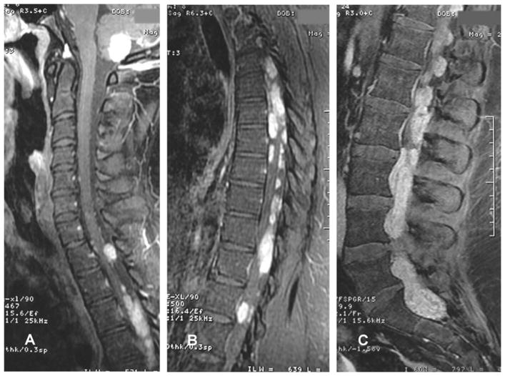
Fig. 1.
Sagittal T1-weighted MRI with contrast demonstrating (A) an expansive, contrast-enhancing intra-axial lesion in the cerebellum with extension into the foramen magnum, and (A, B, C) multiple well-circumscribed enhancing intradural and extramedullary lesions predominantly along the thoracic and lumbar regions. From 85 Macedo LT, Rogerio F, Pereira EB, et al. Cerebrospinal tumor dissemination in a patient with myxopapillary ependymoma. J Clin Oncol 29:2011 e795–798. Reprinted with permission. © 2011 American Society of Clinical Oncology. All rights reserved.

