Figure 1.
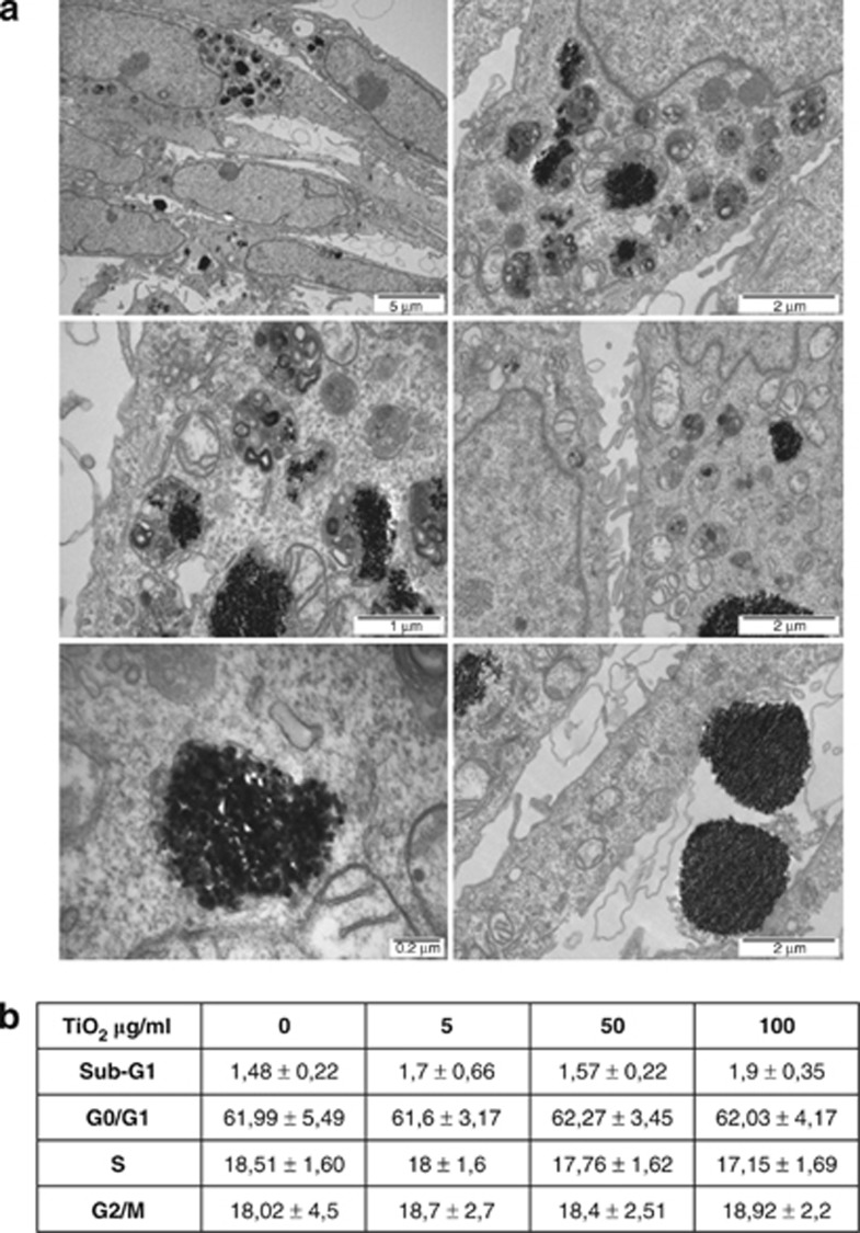
Effect of TiO2 on morphology and cell cycle phase distribution. (a) Transmission electron microscopy of HaCaT cells treated with 50 μg/ml TiO2 for 24 h. Agglomerated and single particles were found inside phagosomes/lysosomes. No particles were found within the nucleus or any other organelles. (b) Apoptosis and cell cycle analysis was performed by flow cytometry in HaCat cells treated for 24 h with different concentrations of TiO2 as indicated. Data represent mean±S.E.M. of three independent experiments

