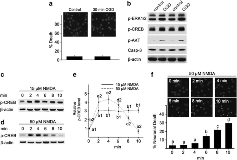
Figure 7.
Short-term insults with OGD or high-dose NMDA do not cause cell death, but rather stimulate the pro-survival signaling. DIV 14 neurons were subjected to a 30-min OGD (a and b), or treated with 15 (c and e) or 50 μM NMDA (d and e) for 2, 4, 6, 8, and 10 min. Samples were collected immediately after the treatment, and examined for the level of p-ERK1/2, p-CREB, p-AKT, and activated caspase-3 (b), or p-CREB (c and d). Quantification of p-CREB after NMDA treatment is shown in (e). ANOVA analysis revealed significant difference (P<0.05) between different groups. The letters a1 and b1 (a1<b1) are different SNK groups for the 15-μM NMDA treatment. The letters a2–e2 (a2<b2<c2<d2<e2) are different SNK groups for the 50-μM NMDA treatment. (a and f) Neurons were washed with culture medium after OGD or NMDA treatment. The neurons were stained with DAPI 20 h later, and cells with condensed nuclear were counted to determine cell death. One-way ANOVA and the post-hoc SNK analysis revealed significant difference (P<0.05) among different groups with a<b<c<d

