Figure 5.
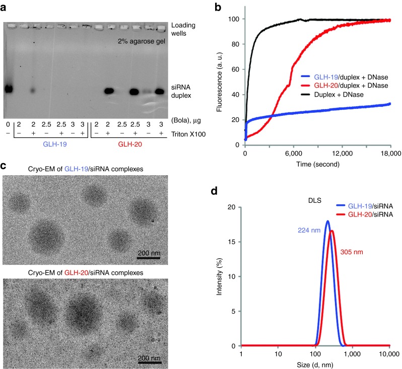
In vitro characterizations of GLH-19/siRNA and GLH-20/siRNA complexes. (a) Formation of complexes was confirmed by agarose gel electrophoresis. siRNA (400 nmol/l final concentration) was mixed with bolaamphiphiles (bola) at final amounts indicated below the gel (in µg). For each amount of bola, equal amounts of detergent Triton X100 were added, aiming to prevent complex formation. Please note that in the case of GLH-19, binding is affected by detergent only at a very low concentration of bola. (b) Relative stabilities of DNA duplexes associated with either GLH-19 (blue line) or GLH-20 (red line) in the presence of DNase. Quenched DNA duplex (100 nmol/l final) labeled with Alexa488 and IowaBlack FQ was mixed with bolaamphiphiles (10 µg final) and DNase was added after 2 minutes of incubation at 37 °C. As the control, naked DNA duplex was completely digested by DNase (black line). Excitation was set at 460 nm and fluorescence signal (arbitrary units (a. u.)) was measured at 520 nm. Please note that there was no significant degradation observed for GLH-19/DNA after 3 hours of incubation. (c) Cryo-EM images of GLH-19/siRNA and GLH-20/siRNA complexes. (d) Size histograms from dynamic light scattering (DLS) experiments indicate average diameters for GLH-19/siRNA (blue) and GLH-20/siRNA (red) to be 224 and 305 nm respectively.

