Figure 1.

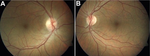
(A) Fundus photograph of the right eye shows swelling of the disc and disc rim hemorrhage (left). (B) Fundus photograph of the left eye shows a healthy appearing but crowded disc with a cup-to-disc ratio of 0.2 (right).

(A) Fundus photograph of the right eye shows swelling of the disc and disc rim hemorrhage (left). (B) Fundus photograph of the left eye shows a healthy appearing but crowded disc with a cup-to-disc ratio of 0.2 (right).