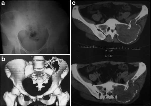
Figure 1.

A 31-year-old woman presented with giant cell tumor involving left sacroiliac joint. Preoperative radiography (a) and computerized tomography (CT) (b) shows an eccentric, geographic, destructive, osteolytic lesion involving the sacrum and posterior superior iliac spine, with slight displacement of the pubic symphysis and left sacroiliac joint (c).
