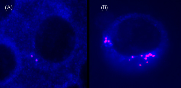
Figure 1.
Different pattern of centrin staining. The isotypic PCs were identified by cytoplasmic or light chain antibody conjugated with AMCA (cIg, blue), and centrin was stained with anticentrin1/2 conjugated with TR. The cells were visualized using Olympus BX-61 fluorescent microscope with Vosskuhler 1300D digital camera and LUCIA-KARYO/FISH/CGH digital analysis system (Laboratory Imaging, s.r.o, Prague, Czech Republic). (For interpretation of the references to color in this figure (A) Two signals – cells with 1–4 signals were considered to have normal centrosome. (B) Abnormal PCs with centrosome amplification (>4 fluorescence signals of centrin).

