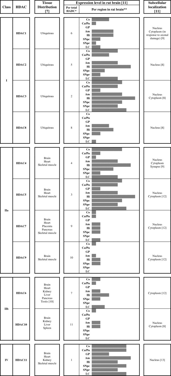
Figure 1.
HDAC isoforms distribution in tissues and rat brain regions, as well as their subcellular localization. Am: Amigadala, As: Astrocytes, Ca/Pu: Caudate/Putamen, Co: Cortex, GP: Globus palidus, Hi: Hippocampus, LC: Locus coeruleus, Ne: neurons, Ol: oligodendrocytes, SNpc: Substantia nigra compacta, SNpr: Substantia nigra reticulata, VEC: Vessel endothelial cells; * classified from 1 (most expressed HDAC isoform) to 11 (less expressed HDAC isoform); ** diagrams are a graphical representation of the relative expression of each HDAC isoform in a scale from low to high (0–5), adapted from Broide et al. [11-13].

