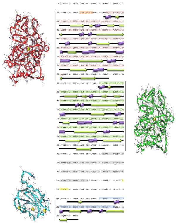
Figure 2.
HDAC6 domain organization. Catalytic Domain I (CDI) primary sequence is highlighted in pale red; CDI three-dimensional structure obtained by homology modeling techniques by using HDAC7 x-ray structure as a template is represented with red ribbons. Catalytic Domain II (CDII) primary sequence is highlighted in pale green; CDII three-dimensional structure obtained by homology modeling techniques by using HDAC7 x-ray structure as a template is represented with green ribbons. Primary sequence of HDAC6 ubiquitin binding domain (ZnFUBP) is highlighted in blue whereas its three-dimensional structure (PDB ID 3C5K) is represented with cyan ribbons. Sequences corresponding to the tetradecapeptide repeating domain (SE14) and to the nuclear export domains NES1, NES2 are highlighted in pale gray, orange and yellow, respectively. Information about HDAC6 CD I/II and ZnFUBP secondary structures was retrieved from the human HDAC7 (PDB ID 3C10) and HDAC6 (PDB ID 3C5K) x-ray structures.

